Why Do Bruises Change from Purple to Yellow? The Chemistry of Hemoglobin Breakdown [Bit#175]
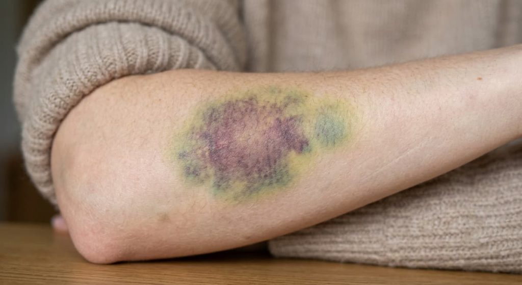
A bruise can look almost purple or blue shortly after an injury, then gradually develop greenish and yellow tones before disappearing. It may seem like the skin is simply changing color as the injury heals, but something much more specific is happening beneath the surface.
The changing colors are closely connected to what happens to hemoglobin after blood escapes from damaged vessels.
Blood Has Left the Vessels
A bruise, or contusion, forms when small blood vessels are damaged and blood leaks into surrounding tissue without breaking the skin.
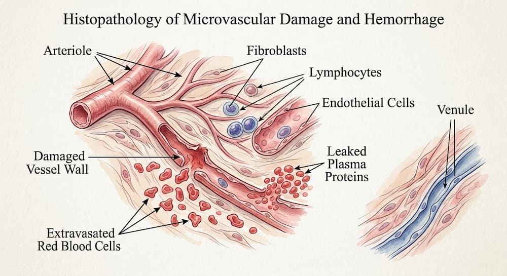
The red blood cells that escape with the blood eventually become trapped outside the circulation. As the tissue responds to the injury, immune cells called macrophages are recruited to the area. These cells help remove damaged red blood cells and process their contents.
This is where the chemistry of the bruise begins to change.
Hemoglobin is the protein inside red blood cells that normally carries oxygen. Its color depends partly on its chemical state and its environment. When blood remains inside the circulation, hemoglobin is continuously maintained within red blood cells and transported through the body. Inside a bruise, however, the blood is isolated from normal circulation and the red blood cells are gradually broken down.
As a result, the pigments associated with hemoglobin begin to change.
From Hemoglobin to Biliverdin
One of the most important components of hemoglobin is heme, a ring-shaped molecule containing iron.
When macrophages break down hemoglobin, the heme portion is processed by an enzyme called heme oxygenase. This reaction converts heme into biliverdin, while also releasing iron and carbon monoxide.
Biliverdin is a green pigment.
This is one reason why a healing bruise can develop a greenish color. However, biliverdin does not simply accumulate indefinitely. In normal conditions, it is rapidly converted into another pigment called bilirubin by the enzyme biliverdin reductase.
The pathway can therefore be simplified as:
Hemoglobin → heme → biliverdin → bilirubin
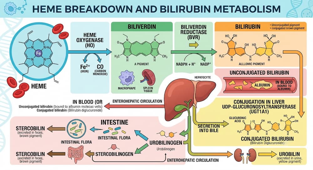
Each step changes the chemical structure of the pigment, and those changes can affect the color visible through the skin.
Why Does the Bruise Become Yellow?
Bilirubin is a yellow-orange pigment best known for its role in normal heme metabolism. The same biochemical pathway that processes old red blood cells throughout the body also helps explain the later stages of a bruise.
As hemoglobin is broken down within the injured tissue, bilirubin and other degradation products accumulate locally. Their presence contributes to the yellow appearance that often develops as a bruise heals.
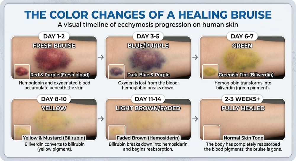
Human studies using optical measurements have found that the yellow component of a bruise is associated with local hemoglobin breakdown. In other words, the yellow color is not simply a sign that the skin is “getting better.” It reflects a chemical transformation of blood-derived pigments within the tissue.
Why Does a Bruise Often Look Purple First?
The early color of a bruise is more complicated than a single pigment.
Freshly leaked blood and hemoglobin within the injured tissue contribute strongly to the initial red, blue, or purple appearance. The amount and location of blood, its oxygenation state, how deeply it lies beneath the skin, and how light is scattered and absorbed by tissue can all influence what the eye sees.
This is why two bruises caused by similar injuries do not necessarily follow exactly the same color sequence.
As the blood is broken down and cleared, the relative contribution of different pigments changes. A bruise may therefore pass through combinations of red, blue, purple, green, brown, and yellow rather than following a perfectly predictable sequence.
The Color Change Is a Chemical Process
The familiar progression of a bruise is therefore a visible consequence of several biological processes happening at once.
First, blood escapes from damaged vessels. Red blood cells then break down outside the circulation. Macrophages remove and process the cellular debris, while enzymes such as heme oxygenase begin dismantling heme.
The resulting pigments change as the molecules are chemically transformed:
Heme → biliverdin → bilirubin
The colors we see are essentially a visual record of this molecular processing, combined with the way light interacts with blood pigments and surrounding tissue.
The process also explains why a bruise does not simply remain purple until it disappears. The material responsible for its appearance is being chemically modified and gradually removed.
Can You Tell How Old a Bruise Is by Its Color?
Not precisely.
Because bruises change color during healing, their appearance has sometimes been used in forensic investigations to estimate how long an injury has been present. But the timing of these changes varies considerably.
A study of 89 people and 369 bruises found that the development of yellow coloration was the most useful visible change associated with time. However, the researchers concluded that the presence of yellow color could indicate that a bruise was more than 18 hours old, rather than providing a precise age.
Factors such as the location of the injury, depth of bleeding, individual differences, and the biological response of the tissue can all affect how a bruise looks.
So a yellow bruise should not be treated as a biological clock.
A Bruise Is More Than a Mark on the Skin
The next time a bruise changes from purple to yellow, what you are seeing is not simply a change in skin color.
It is the visible consequence of a cleanup process happening inside the tissue.
Blood that once carried oxygen through the circulation has escaped from its normal compartment. Red blood cells are dismantled, hemoglobin is processed, heme is converted into biliverdin and then bilirubin, and the resulting products are gradually cleared.
A bruise therefore offers an unusual glimpse of chemistry that normally happens invisibly inside the body: the controlled breakdown of one of the most important molecules in blood.
Scientific Sources
1. Hughes VK, Ellis PS, Burt T, Langlois NEI.
The practical application of reflectance spectrophotometry for the demonstration of haemoglobin and its degradation in bruises.
J Clin Pathol. 2004;57(4):355–359.
PMID: 15047735.
DOI: 10.1136/jcp.2003.011445.
Read the study on PubMed
2. Langlois NEI, Olds K, Ross C, Byard RW.
Heme oxygenase-1 and heme oxygenase-2 expression in bruises.
Forensic Sci Med Pathol. 2015;11(4):482–487.
PMID: 25772118.
DOI: 10.1007/s12024-015-9660-1.
Read the study on PubMed
3. Langlois NE, Gresham GA.
The ageing of bruises: a review and study of the colour changes with time.
Forensic Sci Int. 1991;50(2):227–238.
PMID: 1748358.
DOI: 10.1016/0379-0738(91)90154-B.
Read the study on PubMed
![[Bit#50] The Great Survival Strategy of Plant Hormones](https://hiddenbitknowledge.com/wp-content/uploads/2026/06/64472267701f-768x419.jpg)
![[Bit#49] The Miraculous Defense System of Human Skin](https://hiddenbitknowledge.com/wp-content/uploads/2026/06/1e35ee73e983-768x538.jpg)
![[Bit#111] Human Sixth Sense: Science of Proprioception and Interoception](https://hiddenbitknowledge.com/wp-content/uploads/2026/08/main.six-senses-768x419.jpg)
![[Bit #155] The Secret of Unihemispheric Sleep: Why Some Animals Sleep with Half Their Brains](https://hiddenbitknowledge.com/wp-content/uploads/2026/09/mian.sleeping-768x419.jpg)
![[Bit#167] Why Does the Heart Never Get Tired Even After Beating for a Lifetime?](https://hiddenbitknowledge.com/wp-content/uploads/2026/09/main.heart_-768x384.jpg)
![[Bit#144] Hidden Mechanisms of How Fine Dust Destroys the Lungs](https://hiddenbitknowledge.com/wp-content/uploads/2026/09/main.dust_-767x428.jpg)