What Do Ligaments Do in Your Body? [Bit#202]
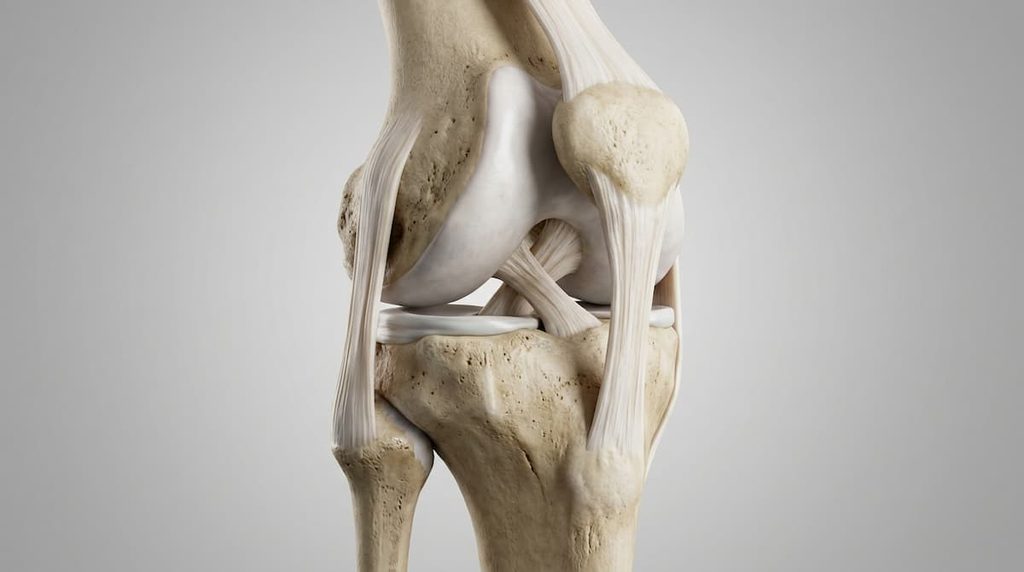
Imagine twisting your knee while playing sports or stepping awkwardly off a curb. Your bones do not simply move in any direction they want. Something helps keep the joints connected and prevents excessive movement.
That something includes ligaments.
Ligaments are strong bands of connective tissue that connect one bone to another. They help hold joints together while allowing them to move. Without them, the bones in your knees, ankles, wrists, and other joints would have much less support.
But not all ligaments do the same job.
Some help prevent the knee from sliding too far forward or backward. Others keep the ankle from rolling too far inward. In the wrist, small ligaments coordinate the movements of tiny bones. Along the spine, several ligaments help control bending and stretching.
So why does the body need so many different kinds of ligaments? The answer becomes clearer when we look at how different joints move.
Ligaments Are Not the Same as Tendons
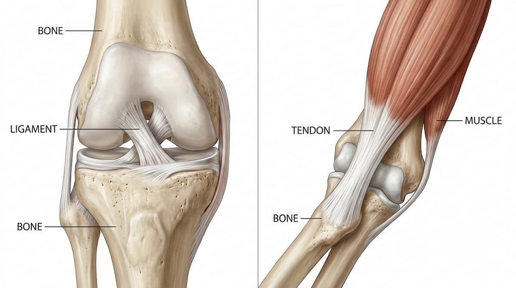
Ligaments and tendons may look similar because both are made of tough connective tissue. However, they connect different structures.
A ligament connects bone to bone, helping stabilize a joint. A tendon connects muscle to bone, transferring the force created when a muscle contracts.
Think about bending your knee. Muscles generate the force needed to move the leg, and tendons help transfer that force to the bones. Meanwhile, ligaments help keep the bones in the correct relationship as the knee moves.
Most ligaments contain large amounts of collagen, a protein that gives connective tissue much of its strength. Collagen fibers are arranged in organized structures that help the tissue resist pulling forces.
However, ligaments are not completely rigid. They can stretch slightly under normal loads, allowing joints to move. Their strength and flexibility help create a balance between movement and stability.
This balance is important because a joint that cannot move would be just as impractical as one that moves too freely.
The Knee Has Ligaments That Control Different Directions
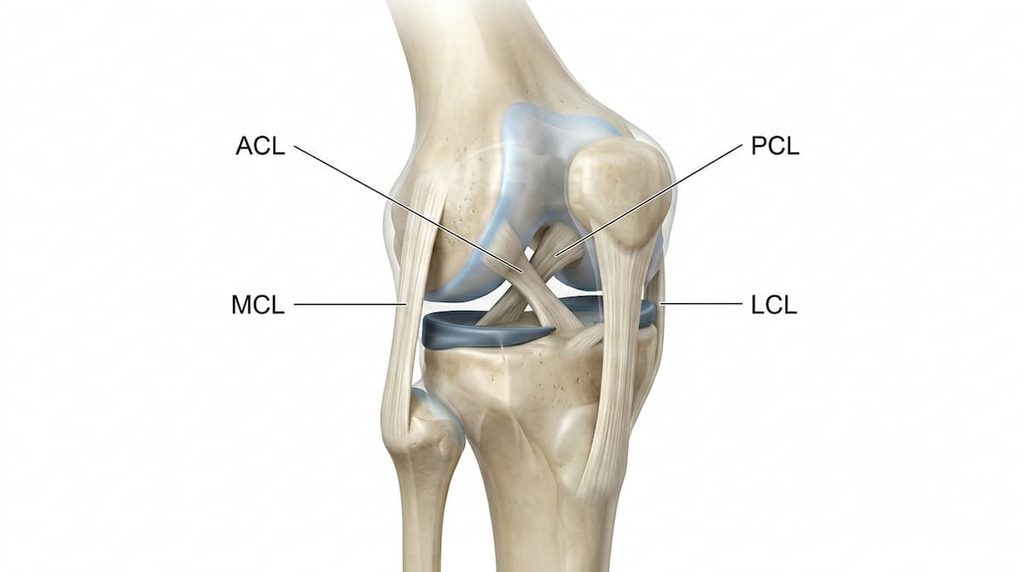
The knee is one of the best examples of how several ligaments work together.
It must support body weight, bend and straighten, and handle forces when we walk, run, jump, or change direction. At the same time, it must prevent excessive movement between the thigh bone and shin bone.
Four major ligaments help provide this stability.
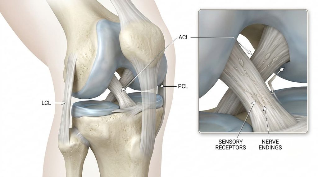
The anterior cruciate ligament, or ACL, sits inside the knee and crosses the posterior cruciate ligament, or PCL. Together, these are called the cruciate ligaments.
The ACL helps resist the shin bone moving too far forward relative to the thigh bone. It also contributes to controlling rotation within the knee.
The PCL performs a different job. It is an important restraint against the shin bone moving too far backward relative to the thigh bone.
The other two major ligaments sit along the sides of the knee.
The medial collateral ligament, or MCL, runs along the inner side. It helps resist forces that push the knee inward. The lateral collateral ligament, or LCL, runs along the outer side and helps resist forces that push the knee outward.
These ligaments do not work independently. Their contributions change depending on knee position and the direction of the force being applied.
This is why a knee injury can affect more than one ligament. A strong twisting force, for example, may place stress on several structures at once.
The knee therefore relies on a coordinated system rather than a single strong band of tissue.
Why Is the Ankle So Easy to Twist?
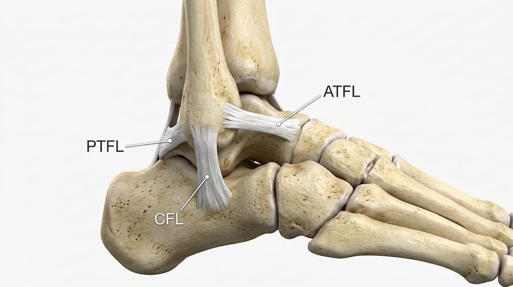
An ankle can roll unexpectedly when you land on an uneven surface or step on the edge of a curb. This movement can stretch or tear the ligaments that support the joint.
The outer side of the ankle has three major ligaments: the anterior talofibular ligament (ATFL), calcaneofibular ligament (CFL), and posterior talofibular ligament (PTFL).
Each has a different position and contribution to ankle stability.
The ATFL connects the fibula, one of the lower leg bones, to the talus in the ankle. It is commonly injured during ankle sprains, particularly when the foot turns inward while pointed downward.
The CFL connects the fibula to the heel bone, called the calcaneus. Unlike the ATFL, it crosses both the ankle joint and the subtalar joint, which lies between the talus and calcaneus.
This means the CFL contributes to stability across more than one joint. It helps resist excessive inward rolling of the foot, although its contribution changes with ankle position.
The PTFL connects the fibula to the back of the talus. It is generally the strongest of the three outer ankle ligaments and is less commonly injured than the ATFL.
There are also important ligaments on the inner side of the ankle. The deltoid ligament complex helps stabilize this side of the joint and resists excessive outward movement of the ankle.
Together, these structures allow the ankle to remain stable while still adapting to uneven ground.
Interestingly, ankle stability does not come from ligaments alone. The shape of the bones, joint surfaces, and surrounding muscles also contribute.
That is why recovering from an ankle sprain involves more than simply waiting for pain to disappear. The joint must regain its ability to move and remain stable during everyday activities.
Small Wrist Ligaments Keep Tiny Bones Working Together
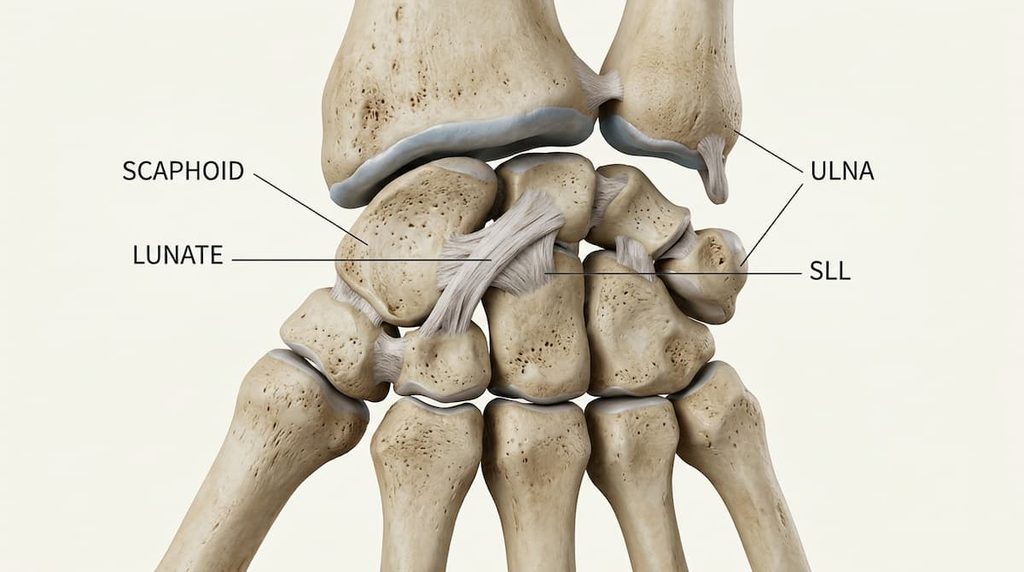
The wrist may look simple from the outside, but it contains many small bones arranged in a complex structure.
These bones need to move together in a coordinated way. If they move too far apart or lose their normal alignment, the wrist can become unstable.
One important structure is the scapholunate ligament.
This ligament connects two wrist bones called the scaphoid and lunate. These bones move in slightly different ways as the wrist bends and rotates.
The scapholunate ligament helps coordinate their movements, allowing them to maintain a stable relationship while the wrist changes position.
Why does this matter?
Imagine trying to grip a heavy object or push yourself up from a chair. Your wrist must transfer forces between your hand and forearm while maintaining its structure.
If the scapholunate ligament is seriously damaged, the scaphoid and lunate may no longer move together properly. This can create instability and, in some cases, contribute to long-term joint damage.
The wrist also contains other important ligaments, including the lunotriquetral ligament, which connects two different wrist bones.
These smaller structures show that ligaments are not only important in large joints such as the knee. Even tiny connections between bones can make a major difference to how a joint works.
The Spine Uses Ligaments to Control Bending
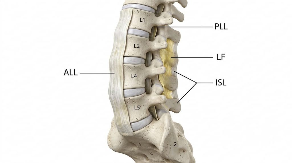
The spine has a very different job from the knee, ankle, or wrist.
It must support the body, protect the spinal cord, and allow the trunk to bend, straighten, and rotate. To accomplish this, it relies on multiple ligaments arranged along and between the vertebrae.
One major structure is the anterior longitudinal ligament. It runs along the front of the vertebral bodies and helps limit excessive backward bending of the spine.
On the opposite side, the posterior longitudinal ligament runs along the back of the vertebral bodies, inside the spinal canal. It helps resist excessive forward bending.
Other ligaments connect the bony projections of neighboring vertebrae.
The supraspinous ligament runs along the tips of the spinous processes, while the interspinous ligaments lie between them. Both become stretched during forward bending and help resist excessive movement in that direction.
Another structure, the ligamentum flavum, connects the laminae of neighboring vertebrae. It contains a relatively high proportion of elastic fibers compared with many other spinal ligaments.
Its elastic properties help it change length as the spine moves and return toward its resting configuration.
These ligaments do not make the spine rigid. Instead, they help control its movement while working together with muscles, joints, and other supporting tissues.
The result is a structure that can remain flexible without allowing every vertebra to move without limits.
Ligaments Do More Than Hold Bones Together
So far, we have seen how ligaments help control the movement of different joints. But there is another interesting part of their function.
Some ligaments also contain sensory nerve endings called mechanoreceptors.
These structures respond to mechanical changes, such as stretching. Their signals contribute to the nervous system’s information about joint movement and position.
This sense is called proprioception. It helps us understand where parts of our body are and how they are moving without constantly looking at them.
For example, you can usually tell whether your knee is bent or straight even with your eyes closed.
Research on the cruciate ligaments of the knee has identified several types of sensory nerve endings that may contribute to this process.
These signals are not the only source of information about joint position. Muscles, tendons, skin, and other structures also provide sensory input.
Still, the presence of these receptors shows that ligaments are not simply passive ropes holding bones together. They also contribute to the sensory system that helps coordinate movement.
Why Every Joint Needs a Different Kind of Support
The knee, ankle, wrist, and spine all contain ligaments, but their requirements are very different.
The knee needs to control forward, backward, and sideways movement while supporting body weight. The ankle needs to remain stable as the foot adapts to different surfaces. The wrist needs small bones to move together without losing their alignment. The spine needs to bend and rotate while limiting excessive movement between vertebrae.
Their ligaments are arranged to meet these different demands.
Yet they share one important purpose. They help joints move within a safe and functional range while maintaining the relationships between bones.
This is also why ligament injuries can have different consequences depending on the joint and the specific structure involved.
A damaged knee ligament may affect stability during running or turning. An injured ankle ligament may make the foot feel unstable on uneven ground. A wrist ligament injury can interfere with gripping and putting weight through the hand.
Ligaments may be small compared with the bones and muscles around them, but their contribution to movement is enormous.
The next time you bend your knee, take a step, or turn your wrist, remember that movement is not simply about muscles pulling on bones. It also depends on the structures that guide and limit that movement.
If every joint moves differently, what would happen if all our ligaments were built exactly the same way?
Scientific Sources
1. Hassebrock JD, Gulbrandsen MT, Asprey WL, Makovicka JL, Chhabra A. (2020). “Knee Ligament Anatomy and Biomechanics.” Sports Medicine and Arthroscopy Review, 28(3), 80–86. PMID: 32740458. DOI: 10.1097/JSA.0000000000000279. Read the study on PubMed
2. Medina McKeon JM, Hoch MC. (2019). “The Ankle-Joint Complex: A Kinesiologic Approach to Lateral Ankle Sprains.” Journal of Athletic Training, 54(6), 589–602. PMID: 31184957. DOI: 10.4085/1062-6050-472-17. Read the study on PubMed
3. Li L, Gollhofer A, Lohrer H, Dorn-Lange N, Bonsignore G, Gehring D. (2019). “Function of ankle ligaments for subtalar and talocrural joint stability during an inversion movement: An in vitro study.” Journal of Foot and Ankle Research, 12, 16. PMID: 30923576. DOI: 10.1186/s13047-019-0330-5. Read the study on PubMed
4. Gopinath R, Pigott M, Lindsey B, Finney FT, Holmes JR, Walton DM, Talusan PG. (2022). “Medial Ankle Instability: Review of Anatomy, Evaluation, and Treatment.” Foot & Ankle Specialist, 15(6), 573–578. PMID: 36210762. DOI: 10.1177/1938640021992915. Read the study on PubMed
5. Rajan PV, Day CS. (2015). “Scapholunate Interosseous Ligament Anatomy and Biomechanics.” The Journal of Hand Surgery, 40(8), 1692–1702. PMID: 26143029. DOI: 10.1016/j.jhsa.2015.03.032. Read the study on PubMed
6. Kapetanakis S, Chatzivasiliadis M, Gkantsinikoudis N. (2025). “Anatomy and biomechanics of posterior ligamentous complex of the human spine: a narrative review.” Surgical and Radiologic Anatomy, 47(1), 170. PMID: 40615623. DOI: 10.1007/s00276-025-03679-9. Read the study on PubMed
7. Panjabi MM, Goel VK, Takata K. (1982). “Physiologic strains in the lumbar spinal ligaments. An in vitro biomechanical study.” Spine, 7(3), 192–203. PMID: 6214027. DOI: 10.1097/00007632-198205000-00003. Read the study on PubMed
8. Banios K, Raoulis V, Fyllos A, Chytas D, Mitrousias V, Zibis A. (2022). “Anterior and Posterior Cruciate Ligaments Mechanoreceptors: A Review of Basic Science.” Diagnostics, 12(2), 331. PMID: 35204424. DOI: 10.3390/diagnostics12020331. Read the study on PubMed
9. Eisner LE, Rosario R, Andarawis-Puri N, Arruda EM. (2022). “The Role of the Non-Collagenous Extracellular Matrix in Tendon and Ligament Mechanical Behavior: A Review.” Journal of Biomechanical Engineering, 144(5), 050801. PMID: 34802057. DOI: 10.1115/1.4053086. Read the study on PubMed