Cut a Planarian, and It Can Regrow Its Body. How? [Bit#178]
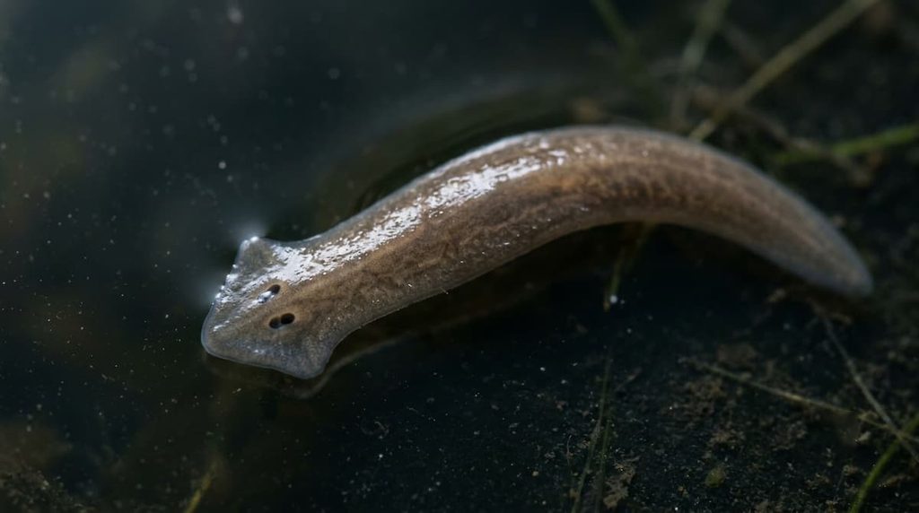
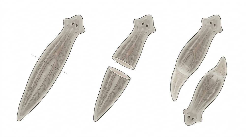
A planarian can lose its head and grow another one. Depending on the species and the piece of tissue that remains, a fragment of this tiny flatworm may rebuild an entire body.
The remarkable part is not simply that new tissue grows. The animal must reconstruct the right structures in the right places. A new head needs eyespots and nervous tissue. A tail fragment must restore its missing front end. The rebuilt animal must recover an organized body rather than becoming an arbitrary collection of cells.
How can a piece of an animal rebuild a structure that is no longer there?
Scientists have found that planarians combine two extraordinary biological abilities: stem cells that can produce many kinds of replacement cells, and molecular signals that help those cells rebuild the correct body pattern. Experiments have even shown that disrupting some of these signals can cause a regenerating fragment to develop a head where a tail should have formed.
The secret is not a single miracle cell. It is a coordinated system for rebuilding a body.
A Tiny Flatworm With an Extraordinary Ability
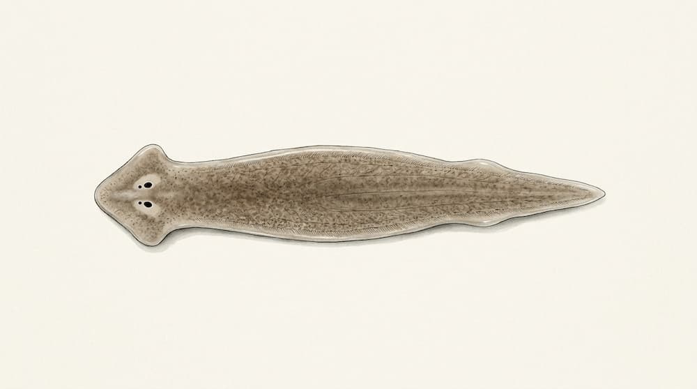
Planarians are free-living flatworms found in freshwater and other aquatic environments. Many species have flattened bodies and two small eyespots near the head. Their simple appearance hides a remarkably flexible system for maintaining and rebuilding tissues.
Regeneration is different from ordinary wound healing. When human skin is injured, cells can close the wound and repair damaged tissue. But human skin does not normally reconstruct an entire missing finger, including its bones, nerves, blood vessels, and joints.
Planarians can solve a much larger problem. Depending on the species and the injury, a body fragment may need to replace several missing tissues and reconstruct an entire body region.
This does not mean every piece of every planarian will survive and become a complete animal. Regeneration depends on the species, the size and location of the fragment, and the conditions under which the animal is kept.
What makes planarians scientifically valuable is their ability to rebuild complex structures from remaining tissue. That gives researchers a way to investigate a fundamental question: what information tells living cells what to rebuild?
The First Essential Ingredient: Neoblast Stem Cells
Throughout a planarian’s body is a population of adult stem cells called neoblasts. These cells are essential for replacing cells lost during normal tissue maintenance and for producing the new cells required after injury.
Some neoblasts are pluripotent, meaning they can generate many different cell types. Other neoblast populations have more restricted developmental potential.
After a substantial injury, neoblasts contribute to the production of replacement cells. Their descendants can differentiate into specialized cells that rebuild the epidermis, nervous system, muscle, digestive tissues, and other structures.
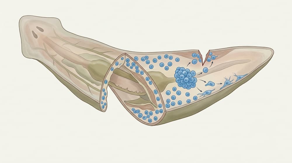
Think of neoblasts as a renewable source of cellular building material. When tissue is missing, the animal needs cells to replace it. Without functional neoblasts, planarians cannot carry out their normal regenerative response.
But there is a problem with this explanation.
Having the right building materials does not tell a cell what to build.
A supply of new cells cannot, by itself, explain why one fragment develops a head while another develops a tail. To solve that problem, planarians need another system that helps organize the new tissue.
What Happens in the First Hours After an Injury?
The response to amputation begins before a new head or tail becomes visible.
Cells around the wound rapidly change their activity. Genes associated with injury responses are switched on, and signaling pathways begin coordinating the response of the remaining tissue.
Studies of planarian regeneration have identified an early, widespread increase in neoblast proliferation at around six hours after injury. A later phase of increased proliferation near the wound becomes prominent by approximately 48 hours when substantial tissue has been lost.
These timings are observations from studied experimental systems, not a universal countdown for every species or injury.
The distinction between minor injury and major tissue loss is important. An incision that removes little or no tissue can trigger an injury response without requiring the reconstruction of an entire body region. A substantial amputation, however, activates additional processes associated with replacing missing tissue.
The animal must do more than close the wound. It must recognize the regenerative challenge, produce the required cells, and establish the correct pattern for the missing structures.
A wound starts the response, but the wound alone does not explain what will grow next.
The Biological Instructions That Tell Cells Where They Belong
Planarians maintain positional information: biological signals and gene-expression patterns that help identify different regions of the body.
This information is essential because the cells at the front of the animal should not behave exactly like cells at the rear. A head and a tail have different structures and functions, so their formation must be controlled differently.
An important discovery is that positional information is not stored exclusively in stem cells. Differentiated cells, especially muscle cells, express genes associated with regional identity. These patterns help the animal maintain its body organization during normal tissue turnover and after injury.
When a fragment loses a major body region, wound-induced signals help re-establish the positional information needed for regeneration.
This provides a solution to the problem of rebuilding missing structures. Neoblasts supply new cells, while positional information helps determine which kinds of cells and structures are appropriate for the region being reconstructed.
The two systems depend on one another. Stem cells without positional instructions would not explain the correct arrangement of the new body. Positional instructions without a source of replacement cells would not be enough to rebuild missing tissue.
Regeneration works because cell production and body-pattern information are coordinated.
The Head-or-Tail Switch: What Wnt Signaling Reveals
One of the most striking discoveries in planarian biology concerns the Wnt signaling pathway.
Wnt signaling helps regulate development and tissue organization in many animals. In planarians, canonical Wnt signaling involving β-catenin plays a major role in establishing posterior identity — the identity associated with the tail end of the body.
The opposite end must develop a head. One molecule involved in this process is Notum, which inhibits Wnt signaling and helps support head regeneration.
These are not merely correlations between molecules and body regions. Researchers have experimentally altered the pathway and observed changes in the structures that regenerate.
When β-catenin signaling is disrupted, a fragment that would normally regenerate a tail can instead develop a head at that end. Depending on the experimental manipulation, the result can be a two-headed planarian.
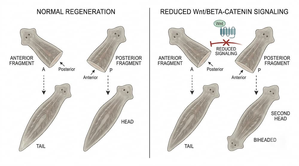
This is a powerful clue. The cells at the wound have not suddenly acquired an entirely new set of physical materials. Instead, changing the signals that regulate body identity changes the structure the tissue develops.
The experiment demonstrates that the identity of a regenerating body region depends on biological instructions, not just on the presence of stem cells.
Wnt signaling is not a universal on-off switch for every aspect of regeneration. Other genes, signaling pathways, and interactions between tissues also contribute. Nevertheless, the Wnt pathway provides one of the clearest examples of how scientists can connect a molecular mechanism to a visible change in body structure.
Why Doesn’t Every Wound Produce a Head?
If Wnt signaling influences whether a head or a tail forms, why does a normal planarian not regenerate two heads every time it is cut?
The answer involves the way several signals interact with the existing body.
In an intact planarian, Wnt-related signals are associated with posterior identity, while Wnt inhibitors such as Notum contribute to anterior identity. After injury, these patterns are modified by wound-induced signals.
Notum is preferentially activated at head-facing wounds, helping establish conditions that support head regeneration. At posterior-facing wounds, Wnt-related signaling helps promote tail identity.
The outcome therefore depends on the orientation of the wound and the signaling environment of the remaining tissue.
Researchers are still investigating how these signals interact with one another and with positional information in different regions of the body. The process cannot be reduced to a single molecule acting independently.
The important point is that regeneration is controlled. A planarian does not simply grow whatever tissue is easiest to produce. It rebuilds structures according to a regulated biological pattern.
Could Electrical Signals Also Influence Regeneration?
Molecular signaling is not the only mechanism scientists have investigated.
Cells also maintain electrical differences across their membranes through the movement of ions. These electrical properties can influence cellular behavior, and researchers have asked whether they help determine the body pattern that develops after an injury.
A 2019 study published in Biophysical Journal investigated early bioelectric signaling in planarians. The researchers found that experimentally altering the electrical state of injured tissue during the first hours after amputation could change subsequent gene-expression patterns and produce two-headed animals under the tested conditions.
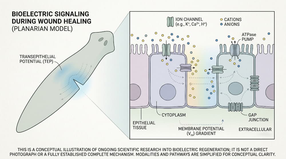
The result suggests that electrical properties can influence the early events that establish head-tail polarity. In other words, the signals that help organize a regenerating body may begin acting before the new structures are visible.
This does not mean scientists have discovered a simple electrical command that can reliably make any animal regrow any body part. The findings concern specific experimental manipulations in a particular model organism.
Instead, they point to a broader possibility: regeneration may depend on several interacting layers of control, including electrical states, molecular signaling, stem-cell behavior, and positional information.
How these layers interact remains an active area of research.
Regeneration Is More Than Growing New Cells
A planarian must do more than replace missing tissue. It must restore the correct arrangement of structures and maintain that organization after regeneration is complete.
This is closely related to tissue homeostasis, the process by which an organism maintains its tissues during everyday life. Even without a major injury, planarians continually replace cells. Neoblasts contribute to this ongoing renewal, while positional information helps preserve the organization of the body.
Regeneration draws on the same broad biological systems but places them under a different demand: instead of replacing individual cells or small amounts of tissue, the animal must rebuild structures that have been removed.
Scientists continue to investigate how the animal coordinates cell production, tissue patterning, and the restoration of a stable body. The precise mechanisms that determine when every structure has been correctly restored are not fully understood.
This is one reason planarians remain useful research organisms. They allow scientists to study how stem cells and positional signals work together, rather than examining either process in isolation.
What This Means for Human Regeneration
Planarian research has implications beyond flatworms because many biological signaling systems are shared across animal species.
Humans also rely on stem cells and molecular pathways to maintain and repair tissues. Studying planarians can therefore help researchers understand general principles of cell differentiation, tissue organization, and responses to injury.
But there is a crucial limitation.
A pathway that helps a flatworm rebuild its head does not mean the same pathway can make a human regrow an entire organ.
Human tissues have different structures, developmental constraints, and regenerative capacities. Similar molecules can play different roles depending on the tissue and the organism. Findings in planarians are valuable for understanding biology, but they do not provide a direct recipe for human limb or organ regeneration.
The long-term value of this research is more fundamental. It helps reveal how living systems coordinate the production of cells with the information needed to organize those cells into functional tissues.
Planarians demonstrate that rebuilding a body requires more than a powerful population of stem cells. The cells must receive and respond to the right instructions.
Their regeneration is the result of a coordinated system: stem cells provide the new tissue, positional information guides its organization, and signaling pathways help determine which structures form at each end of the body.
Understanding how these systems work together may help scientists discover new approaches to tissue repair — even if the extraordinary regenerative abilities of planarians remain far beyond those of humans.
Scientific Sources
- Reddien, Peter W. The Cellular and Molecular Basis for Planarian Regeneration. Cell, 2018, 175(2), 327–345. DOI: 10.1016/j.cell.2018.09.021. PMID: 30290140. Read the study on PubMed
- Petersen, Christian P., and Peter W. Reddien. A wound-induced Wnt expression program controls planarian regeneration polarity. Proceedings of the National Academy of Sciences, 2009, 106(40), 17061–17066. DOI: 10.1073/pnas.0906823106. PMID: 19805089. Read the study on PubMed
- Petersen, Christian P., and Peter W. Reddien. Polarized notum activation at wounds inhibits Wnt function to promote planarian head regeneration. Science, 2011, 332(6031), 852–855. DOI: 10.1126/science.1202143. PMID: 21566195. Read the study on PubMed
- Reddien, Peter W. Positional Information and Stem Cells Combine to Result in Planarian Regeneration. Cold Spring Harbor Perspectives in Biology, 2022, 14(4), a040717. DOI: 10.1101/cshperspect.a040717. PMID: 34518341. Read the study on PubMed
- The Role of Early Bioelectric Signals in the Regeneration of Planarian Anterior/Posterior Polarity. Biophysical Journal, 2019, 116(5), 948–961. DOI: 10.1016/j.bpj.2019.01.029. PMID: 30799071. Read the study on PubMed
![[Bit#21] Cockroaches: The Ultimate Biological Design Surpassing Nuclear Survival](https://hiddenbitknowledge.com/wp-content/uploads/2026/05/015d9c51b899-768x512.jpg)
![[Bit#78] The Perfect Predator: The 300 Million Years Secret Hidden by Dragonflies](https://hiddenbitknowledge.com/wp-content/uploads/2026/06/main.dragonfly-768x512.jpg)
![[Bit#115] The Hidden Secrets of Seahorses: Nature’s Most Unusual Marine Creatures](https://hiddenbitknowledge.com/wp-content/uploads/2026/08/main.Seahorse-768x513.jpg)
![[Bit#126] Alien Biology Unveiled: What Forms Could Extraterrestrial Life Take Beyond Earth?](https://hiddenbitknowledge.com/wp-content/uploads/2026/08/main.-Alien-Biology-Unveiled-768x419.jpg)
![[Bit#165] Green Sea Slug: The Secret of an Animal Powered by Sunlight Like a Plant](https://hiddenbitknowledge.com/wp-content/uploads/2026/09/main.Elysia-chlorotica-767x428.jpg)
![[Bit#113] Ant Natural Preservatives: Chemical Weapons Defeating Fungi](https://hiddenbitknowledge.com/wp-content/uploads/2026/08/main.ant-macro-768x430.jpg)